

PET CT Imaging: Advanced Support for Cancer Diagnosis and Care
Accurate diagnosis is an essential part of effective cancer management. Modern imaging technologies allow healthcare professionals to examine the body's structures and understand changes in tissue activity. PET and CT technologies can work together to provide valuable information about suspicious areas. This combined approach can support cancer diagnosis, staging, treatment planning, and follow-up when recommended by a medical specialist.
What Is PET CT Imaging?
PET CT imaging is a specialized examination that combines Positron Emission Tomography (PET) with Computed Tomography (CT). Each technology provides different information. CT produces detailed anatomical images of organs and tissues, while PET shows areas of increased metabolic activity.
Before the scan, a small quantity of radioactive tracer is usually administered. The tracer moves through the body and may collect in tissues with higher metabolic activity. The PET scanner detects this activity, while CT images help specialists identify the exact location and structure of the area being evaluated.
How PET CT Supports Cancer Evaluation
Many cancer cells can demonstrate higher metabolic activity than surrounding normal tissues. PET imaging may highlight these areas, while CT helps provide anatomical detail. Combining the two sets of information can give physicians a more complete picture than either examination alone in appropriate clinical situations.
PET CT may be used in the evaluation of several cancers, including certain lung cancers, lymphomas, colorectal cancers, breast cancers, and head and neck cancers. Its usefulness depends on the type of cancer, clinical circumstances, and tracer selected for the examination.
PET CT for Cancer Diagnosis
PET CT for cancer diagnosis can provide useful information when a healthcare professional is investigating a suspicious abnormality or evaluating an already confirmed cancer. It may help identify metabolically active lesions and assess whether abnormalities appear confined to one location or occur in multiple areas.
PET CT results should not be considered separately from other diagnostic information. Depending on the situation, doctors may also consider biopsy findings, laboratory tests, medical history, physical examination, ultrasound, MRI, or other imaging studies before establishing a diagnosis.
PET CT and Cancer Staging
After cancer has been diagnosed, determining its stage is important for treatment planning. Staging provides information about the primary tumor and whether cancer may have involved nearby lymph nodes or distant organs.
PET CT can evaluate multiple regions of the body and may identify areas with abnormal tracer uptake. This can help specialists assess the possible distribution of disease and determine whether additional testing or treatment planning is required.
Monitoring Response to Treatment
PET CT may also be used during cancer treatment and follow-up. Depending on the cancer type and individual treatment plan, doctors may recommend scans at specific intervals to evaluate changes in disease activity.
Comparing current images with earlier examinations can provide information about changes in metabolic activity and anatomy. A decrease in abnormal activity may indicate a response to treatment, while persistent or increased activity may require additional assessment by the treating team.
Preparing for a PET CT Examination
Preparation depends on the type of PET CT study and the tracer being used. Patients may receive instructions regarding food, beverages, physical activity, and medications before the appointment.
Individuals with diabetes or other relevant medical considerations should inform the imaging team before the procedure. Following all preparation instructions carefully can help ensure that the examination is performed appropriately and that useful images are obtained.
What Happens During PET CT?
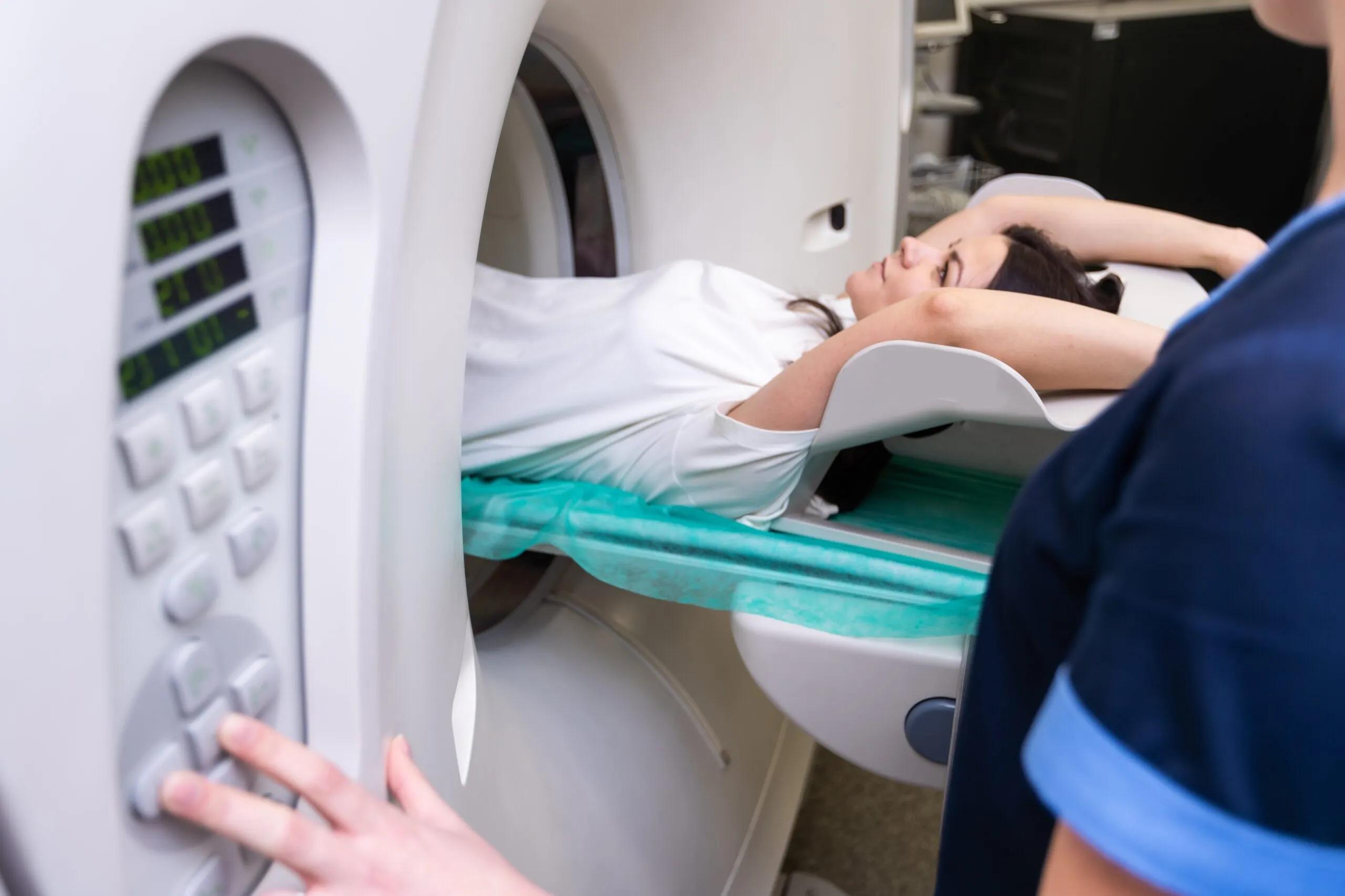
After the tracer is administered, patients usually rest quietly for a specified period. This allows the tracer to distribute throughout the body. During the scan, patients need to remain still so that clear images can be produced.
The imaging team provides instructions throughout the procedure. The examination itself is generally painless apart from the injection, although the complete appointment can take several hours because it includes preparation, tracer uptake, and image acquisition.
Importance of Professional Interpretation
PET CT images require careful interpretation by appropriately trained medical professionals. Specialists consider the patient's symptoms, previous examinations, treatment history, and other clinical information when evaluating the findings.
This comprehensive approach helps doctors understand the significance of imaging results and determine whether additional tests or follow-up may be appropriate.
Conclusion
PET CT is an advanced imaging technique that combines functional and anatomical information to support modern cancer care. It can assist with evaluating suspicious abnormalities, staging confirmed disease, planning treatment, and monitoring response. When recommended by a healthcare professional, PET CT can be an important part of a comprehensive diagnostic and oncology management plan.